
The cardiovascular system plays a vital role in the overall function of the body. It’s responsible for transporting oxygen and nutrients to the heart and organs and maintaining homeostasis. If you’re experiencing symptoms of venous insufficiency or peripheral artery disease, your medical provider may require you to get a vascular ultrasound.
Vascular scans are designed to assess blood flow and detect early signs of vascular diseases. There are a variety of different types of vascular tests. Each ultrasound is designed to evaluate cardiovascular health and help identify the existence of blood clots, hypertension, or other abnormalities.
In this article, we’ll look at the different types of vascular tests and how your medical provider may use these diagnostic tools to provide treatment plans for your condition.
What is a Vascular Ultrasound?
A vascular ultrasound is an advanced imaging technique medical professionals use to assess blood flow in the veins and arteries. These scans help diagnose blood clots, blockages, and other common vascular diseases. With the help of vascular ultrasounds, your medical provider can monitor your blood flow and help you develop a treatment plan for your condition.
Schedule a Treatment Plan Consultation
How is a Vascular Ultrasound Performed?
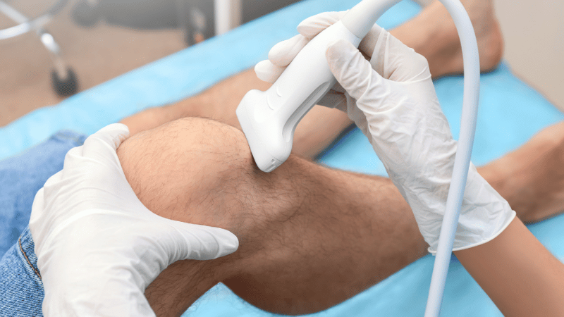
Vascular scans are a non-invasive test that causes minimal to no discomfort. It uses high-frequency sound waves to photograph blood vessels and examine blood flow.
On the day of your ultrasound, a medical professional will apply a lubricant to the skin. They will then place a sonographer on the skin and move it over the specific area. The sonographer takes images of your blood vessels to assess blood flow speed from different angles.
There are a variety of different types of vascular tests. Each test provides insight into your overall circulatory health. In many instances, a medical provider may use various ultrasounds to gain a complete, comprehensive understanding of your vascular health.
Seven Different Types of Vascular Ultrasounds
Depending on your symptoms and overall cardiovascular health, there are several types of vascular tests a medical provider may recommend. Each scan provides insight into your vascular health and can help diagnose various vascular conditions.
1. Arterial Doppler Ultrasound
Arterial Doppler ultrasounds measure blood flow in the arteries. Arteries are designed to move oxygen from the heart towards the organs. This supplies nutrients to the rest of your body, nourishing the organs, tissues, and cells. This vascular scan helps medical professionals better understand blood flow velocity in the arteries, providing a more robust understanding of the overall health of your arteries.
This vascular scan can help identify blood clots, and arteriosclerosis and diagnose peripheral artery disease (PAD). Arterial Doppler ultrasounds are typically performed on the legs, arms, and neck. A medical professional may order this type of ultrasound for patients experiencing stroke symptoms or a decrease in blood flow.
2. Color Doppler Ultrasound
Color Doppler ultrasound uses different colors to assess blood flow and identify abnormalities in both vessels and arteries. If a patient is experiencing swelling in the legs, a medical professional may recommend this ultrasound to check for any aneurysms or blood clots. These vascular tests use color-flowing imaging. The scan will typically use blue, red, and grayscale to highlight different directions of blood flow.
A color Doppler ultrasound is also the main way to test for deep vein thrombosis (DVT). This is a cardiovascular condition where blood clots form in the deep veins, commonly found in the legs. Individuals with DVT may struggle with prolonged sitting, varicose veins, or cramping in the lower extremities.
This vascular scan is different from an arterial Doppler ultrasound. While an arterial Doppler ultrasound focuses primarily on the health of your arteries, a color Doppler ultrasound provides a broader evaluation of blood flow.
3. Venous Doppler Ultrasound
This type of vascular test measures blood flow in the veins. Veins carry deoxygenated blood from the organs and back to the heart. This vascular scan helps medical professionals better understand the speed of blood flow returning to the heart and the overall blood circulation.
Venous Doppler Ultrasounds are commonly performed on the legs and arms but can also be used on other parts of the body where veins exist. A vascular specialist may recommend this vascular scan to patients experiencing swelling, pain, or discoloration in the legs and arms. Early signs of detection of major venous and vascular diseases can help minimize risk and symptoms.
4. Duplex Ultrasound
This type of vascular test evaluates the speed of blood flow. This vascular scan is called a “duplex ultrasound” because it combines two ultrasound techniques: B-mode and Doppler ultrasounds. B-mode ultrasounds are traditional ultrasounds used to take images of your blood vessels. While Doppler ultrasounds assess the speed of blood flow through vessels.
With both vascular scanning techniques, medical professionals can detect narrowing, blood clots, blockages, atherosclerotic plaques, and other abnormalities. Patients with a history of stroke or who show early signs of artery disease may need to undergo this type of vascular test.
5. Triplex Ultrasound
A triplex ultrasound combines three types of image scanning: 2D imaging, color Doppler, and Doppler ultrasound. When all three tests are combined, they provide a comprehensive understanding of blood flow in the arteries and veins.
These vascular tests provide a more detailed understanding of the blood flow patterns in the body and help diagnose carotid artery disease, aneurysms, renal artery stenosis, or chronic venous insufficiency.
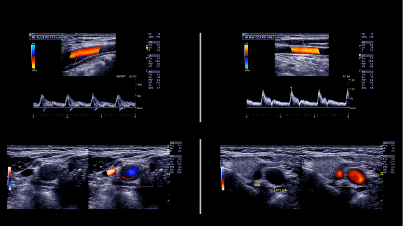
6. Carotid Ultrasound
Commonly referred to as a carotid duplex, this vascular test assesses blood flow in the carotid arteries. These arteries are located on either side of the neck and are vital for supplying blood to the brain. Your medical provider may recommend this ultrasound if you recently had a stroke or exhibited early signs of carotid artery disease.
7. Aortic Ultrasound
An aortic ultrasound is an advanced imaging scan that evaluates the aortic artery. Medical professionals typically use a duplex ultrasound scan to look for aneurysms, ruptures, or infections. Weaknesses in the aorta can be fatal if they aren’t caught early. This ultrasound is a preventative measure that medical professionals typically recommend for patients with specific risk or genetic factors.

Take Control of Your Cardiovascular Health With USA Vascular Centers
At USA Vascular Centers, we offer a variety of minimally invasive, non-surgical treatment plans for common vascular diseases. There are three popular treatments for PAD. This includes lifestyle changes, medication, and a special procedure. Once you schedule a consultation with one of our vascular specialists, our team will help you develop a PAD treatment plan that aligns with your health needs.
We’re here to help you develop a treatment plan that aligns with your cardiovascular needs and gets you one step closer to a healthier you.
