

Medically reviewed by
USA Clinics Medical Review Team
Vascular Health Education Experts
Reviewed: September 2026
Atherectomy removes plaque from a narrowed artery; angioplasty compresses it against the arterial wall. Both are minimally invasive, catheter-based procedures used to treat peripheral artery disease (PAD), and both can be performed on an outpatient basis with no general anesthesia required. The distinction that matters most for patients is not which procedure is inherently superior, but which is better suited to the specific type of blockage and the anatomy of the affected artery. During evaluation, a vascular specialist uses imaging to make that determination.
Understanding how these two approaches differ, and when each is used, can help patients prepare for conversations with their vascular care team and make sense of a recommended treatment plan.
What Is the Main Difference Between Atherectomy and Angioplasty?
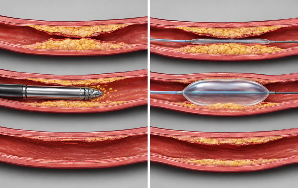
The core distinction between the two procedures comes down to what happens to the plaque itself. Atherectomy physically removes plaque from the artery wall. Angioplasty pushes it out of the way without removing it. Both approaches restore blood flow through a narrowed or blocked artery, but they do so through fundamentally different mechanisms.
How Atherectomy Removes Plaque
During an atherectomy procedure, a vascular specialist guides a specialized catheter to the site of the blockage using real-time imaging. At the tip of the catheter is a cutting or rotating device, depending on the type of atherectomy being performed, that shaves, grinds, or vaporizes plaque directly from the artery wall. The removed material is collected in a small chamber at the catheter tip and withdrawn from the body, clearing the arterial passageway rather than just compressing it. Learn more at the USA Vascular Centers atherectomy procedure page.
How Angioplasty Opens the Artery
Angioplasty, also called percutaneous transluminal angioplasty (PTA), uses a different approach. A catheter with a small, deflated balloon at its tip is guided through the blood vessel to the site of narrowing. The balloon is then inflated, pressing the plaque against the artery wall and widening the interior channel. The balloon is deflated and removed, leaving a wider pathway for blood to flow through. In many cases, a small mesh tube called a stent is placed inside the artery after angioplasty to help hold it open and reduce the chance of re-narrowing.
When Does a Vascular Specialist Recommend Atherectomy?
Atherectomy tends to be the preferred approach when plaque has become heavily calcified, meaning it has hardened over time in a way that makes it resistant to compression from a balloon alone.
Heavily Calcified Blockages
Calcified plaque can have an almost bone-like consistency that does not compress well under balloon pressure. In these cases, attempting angioplasty first may yield incomplete results because the balloon cannot adequately widen the artery. Atherectomy addresses this by removing the hardened material directly, which may allow the artery to open more fully. For patients with leg pain from PAD associated with long-standing or heavily calcified blockages, atherectomy may be evaluated as part of the initial treatment plan.
Atherectomy as Vessel Preparation
One use of atherectomy that is not widely discussed in patient-facing content is its role as a preparation step rather than a standalone procedure. A vascular specialist may perform atherectomy first to break up calcified or complex plaque, then follow it immediately with a drug-coated balloon or stent placement. This staged approach can improve the result of the subsequent angioplasty step by allowing the balloon to work more effectively on a better-prepared artery wall. The decision to combine procedures in this way depends on the extent and type of the blockage, which is evaluated during imaging.
When Is Angioplasty the Right Approach?
Angioplasty tends to work well when the blockage involves softer plaque or when the narrowing is in a location where a stent can be placed safely and effectively.
Softer or Shorter Blockages
When plaque has not calcified extensively, a balloon can often compress it adequately against the artery wall without first needing to remove it. Angioplasty is a well-established technique with high technical success rates for many types of arterial narrowing, and it typically requires a shorter procedure time than atherectomy. Patients exploring treating peripheral artery disease without open intervention often encounter angioplasty as a first-line consideration for moderate, softer blockages.
Angioplasty With Stent Placement
Stent placement frequently follows angioplasty. After the balloon opens the artery, a metal mesh tube is placed inside to act as a scaffold that holds the vessel open over time. This combination may reduce the likelihood of the artery narrowing again in the treated segment. Drug-eluting stents, which release medication into the artery wall, may be used in some cases. The right stent type and placement decision is made by the vascular specialist based on the location and characteristics of the blockage.
Can Atherectomy and Angioplasty Be Used Together?
Yes. In many real-world treatment scenarios, these procedures are used in combination rather than as alternatives to each other. A common approach involves performing atherectomy first to clear or reduce a calcified blockage, then using angioplasty, with or without stenting, to optimize the final result. This is sometimes described as a staged approach.
The availability of both options within a single treatment session gives vascular specialists flexibility to adapt to what they find during the procedure. At USA Vascular Centers, specialists assess each patient’s arterial anatomy using imaging before and during the procedure, selecting the appropriate tools based on what will produce the best outcome for that individual’s specific blockage pattern. Review the full range of PAD treatment options available at USA Vascular Centers to understand how these approaches fit into a broader care plan.
What to Expect During Each Procedure

Both atherectomy and atherectomy procedures follow a similar preparation and recovery framework, though the details of each step differ.
During an Atherectomy
An atherectomy is typically performed under local anesthesia with light sedation, meaning patients remain awake but comfortable. A vascular specialist puts a catheter (a tiny tube) in the groin or upper thigh and guides it through the blood vessel to the site of the blockage. Real-time imaging is used throughout the procedure to ensure accurate placement. The cutting or rotating mechanism at the catheter tip removes the plaque, which is collected and withdrawn. The procedure generally takes less than two hours. After a brief observation period at the outpatient center, most patients are able to return home the same day.
During an Angioplasty
Angioplasty follows a similar setup. After a small access point is created, a balloon catheter is guided to the narrowed segment of the artery under imaging. The balloon is inflated to compress the plaque, then deflated and removed. If a stent is being placed, it is positioned and expanded in the same session before the catheter is withdrawn. Angioplasty typically takes 30 to 90 minutes. Recovery at home for most patients follows the same same-day discharge model as atherectomy. Post-procedure instructions generally include avoiding heavy exertion for a short period and taking any prescribed medications as directed.
How a Vascular Specialist Determines the Right Approach
The choice between atherectomy and angioplasty, or a combination of both, is not made before a thorough evaluation. A vascular specialist begins with a clinical assessment that includes an ankle-brachial index (ABI) test, which compares blood pressure in the ankle and arm to identify reduced arterial flow. Additional imaging, such as vascular ultrasound, may be used to identify where and how severely an artery has narrowed.
The degree of calcification in the plaque, the length and location of the blockage, and the patient’s overall vascular anatomy all factor into the treatment recommendation. Some locations in the leg are treated differently from others based on vessel behavior and the long-term outcomes associated with specific approaches in those areas. A patient’s history with prior interventions may also inform the plan.
Patients who have been experiencing PAD symptoms such as leg cramping during activity, skin changes, or slow-healing sores on the foot should not self-select between procedures. The appropriate first step is a vascular evaluation, where imaging data rather than symptom description guides the treatment decision. To schedule a vascular evaluation at a USA Vascular Centers location, patients can book directly online.
Next step
Understanding your PAD treatment options starts with an evaluation.
A vascular specialist can use imaging to assess your arteries, determine the extent of any blockages, and recommend whether atherectomy, angioplasty, or a combination approach may be appropriate for your situation.
Frequently Asked Questions About Atherectomy and Angioplasty
- National Center for Biotechnology Information. “Peripheral Arterial Disease.” StatPearls. ncbi.nlm.nih.gov/books/NBK430745/
- Jaff MR, et al. “Management of Patients with Peripheral Artery Disease.” Circulation, 2015. pmc.ncbi.nlm.nih.gov
- Shammas NW, et al. “Atherectomy-assisted versus percutaneous angioplasty interventions for treatment of symptomatic infra-inguinal peripheral arterial disease.” PMC6900743. pmc.ncbi.nlm.nih.gov/articles/PMC6900743/
- Botti CF Jr, et al. “Comparison of Atherectomy to Balloon Angioplasty and Stenting for Isolated Femoropopliteal Revascularization.” Journal of Vascular Surgery, 2020. sciencedirect.com
Medical disclaimer: USA Vascular Centers content provides medical education only. It is not a diagnosis or treatment recommendation. Consult a vascular specialist for evaluation.